Sponsored Content | November 06, 2023
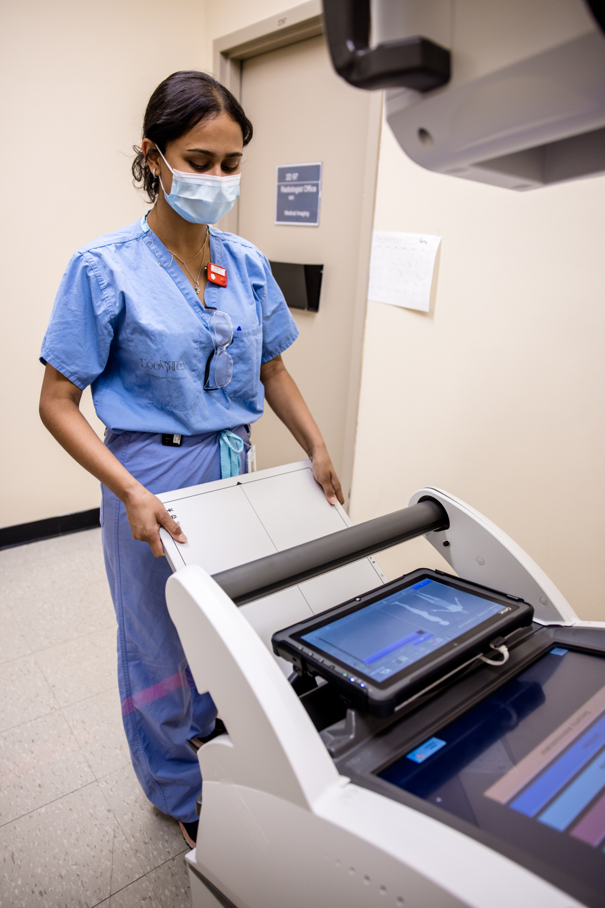
Looking to find an alternative medical imaging option that can be easily implemented and provide extra clinical information for the ICU population at the bedside, KA Imaging and Grand River Hospital (GRH) have partnered on an innovative commercialization project supported by the Coordinated Accessible National (CAN) Health Network. Aiming to transform existing hospital workflow for intensive care unit (ICU) imaging, GRH has added KA Imaging's Reveal 35C, a device that is designed to simultaneously produce both conventional chest X-ray at low dose and higher-contrast spectral radiographic images for improved patient monitoring and faster, more accurate bedside imaging in its ICU.
The hospital has been measuring success metrics around image quality, impact on work processes, and whether follow-up imaging was needed after Reveal's images.
"A preliminary analysis comparing the six weeks preceding the pilot period and the 6 weeks in which KA Imaging's 35C Reveal detector was in use demonstrates a decrease in both the total number of portable chest x-rays as well as chest CTs for patients admitted to ICU. The proportion of all CTs that were chest CTs decreased by approximately 8% and the proportion of patients that required both a chest CT and a portable chest x-ray also decreased by approximately 7%. Though these are early results and further analysis is required, the overall trend is very promising," said Carla Girolametto, Director of Innovation, Research, and Clinical Trials at Grand River Hospital.
ICU: a challenging environment for imaging
Medical imaging plays a critical role in monitoring the condition of ICU patients, commonly to check for conditions like pneumonia or pneumothorax or to rule out other potentially serious pulmonary issues along with verifying the tips of catheters or endotracheal tubes. Imaging in the ICU can be challenging because of reduced patient mobility, the need for imaging outside regular operating hours, and the need for quick imaging turnaround for bedside decision making.
Generally limited on tissue differentiation, portable chest radiography can be ineffective at accurately spotting complex pulmonary issues and sometimes even to localize the tips of lines and tubes. Other modalities like CT are not portable, bring increased radiation exposure, in addition to risks associated with intra-hospital patient transportation. Furthermore, reimbursement for ICU patients is capitated so an unnecessary CT scan increases cost of care and the financial burden for hospitals.
SpectralDR: 3 images, 1 exposure, increased diagnostic information
KA Imaging's Reveal™35C is a single-exposure, portable, digital dual-energy subtraction X-ray detector. It is powered by SpectralDR technology, which produces spectral images that separate materials such as water (like soft tissue, lung lesions) and calcium (such as bones, retained foreign objects, in dwelling devices or other calcifications) and are higher contrast thus, easier to read for a variety of clinicians of varying ability. It also uses the same radiation dose as a traditional X-ray to create the 3 different images without blurring or streaking due to patient movement.
KA Imaging's device has been installed on one of GRH's existing portable x-ray machines and has been piloted to help clinicians validate patient tube and line placements as well as monitor the health of patients to prevent respiratory conditions.
Learn more about SpectralDR technology