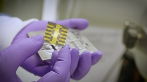
Researchers have developed an X-ray detector out of thin films of perovskite, making it more sensitive and affordable to manufacture than traditional silicon-based detectors.
Researchers in New Mexico and Illinois have prototyped a self-powering X-ray detector from a unique material that enables medical imaging at a fraction of the radiation exposure from conventional X-ray imaging — and a fraction of the manufacturing cost.
The Los Alamos National Laboratory in Santa Fe and Argonne National Laboratory at the University of Chicago swapped out the usual silicon-based technologies used in detectors with a structure built around a thin film of the mineral perovskite. The result, they report, is a one hundred-times more sensitive solution that does not require an outside power source.
“It converts X-ray signal directly into electrical signal, thus offering high sensitivity and in situ real-time X-ray photon tracking capabilities,” Wanyi Nie, a staff scientist at Los Alamos National Laboratory, told HCB News. “The improved detection limit will help to reduce required X-ray dosage to achieve high-quality images and protect patients from being exposed under high dosage. We also expect our technology to provide higher spatial resolution to resolve soft matters and provide more details.”
Rich in heavy elements such as lead and iodine, perovskite can more readily absorb X-rays that pass through silicon undetected, enabling it to better detect high-energy X-rays. This makes it better able to monitor X-rays at high-energy research facilities, such as synchrotron light sources.
Perovskite films can be deposited on surfaces by spraying solutions that cure and leave thin layers of the material behind. This allows for easier and cheaper detector production compared to silicon-based systems, which require high-temperature metal deposition under vacuum conditions. The detectors could potentially be printed large scale with ink-jet systems, replacing half-million-dollar silicon detector arrays.
The direct conversion makes it valuable for real-time imaging and allows for the signal to be directly connected to the computer system for data processing and visualizations, according to Nie.
“There are radiation therapy techniques of using gamma-ray to treat cancers and tumor cells,” Nie said. “I think a detector that can monitor gamma-ray photons in real time would be very useful in those applications. Another advantage of a sensitive solid-state detector is the possibility of resolving radiation photon energy by counting the single radiation photons by electrical pulses, offering more advanced functionalities to medical imaging. For instance, it can differentiate tumors from regular tissues if they absorb different energy photons differently.”
The prototype currently has a single pixel demonstration, with the developers aiming to produce arrays with reduced pixel sizes in order to build a camera device for imaging applications. It expects wide use of the scanner if array testing proves to be successful.
Nie led the project alongside Oppenheimer Distinguished Postdoc Fellow Hsinhan Tsai — both Los Alamos National Laboratory material scientists. The two worked in conjunction with the Los Alamos Theory team led by Sergei Tretiak and Joseph Strzalka at Argonne National Laboratory's X-ray Science Division.