by
Lisa Chamoff, Contributing Reporter | July 01, 2019
From the July 2019 issue of HealthCare Business News magazine
Ikonopedia
Ikonopedia, which offers a cloud-based structured breast reporting system known as the Ikonopedia Breast Reporting and Tracking software, recently released an automated combined reporting feature that automatically combines the diagnostic mammogram and other breast imaging modalities, including ultrasound, MRI and biopsies, into a single report.
The feature is designed to simplify the reporting process, save time and prevent errors, according to Emily Crane, Ikonopedia's chief executive officer.
Part of that same goal is a new feature that is part of the pathology module of the Ikonopedia Breast Reporting and Tracking software, which allows the pathology results from a biopsy to be automatically added to the biopsy or imaging report.
Pathology results are also connected to the radiologist’s areas of concern in the breast.
“Anytime a doctor says, ‘Here’s a place I’m worried about on an image,’ we tie it directly to the finding,” said Crane, who noted that other reporting and tracking software doesn’t go down to a specific area of concern. “Everything we do is at a very specific concern level, instead of a breast level.”
Ikonopedia has also updated its tablet-based patient questionnaire, which employs a web-based version of the Tyrer-Cuzick Breast Cancer risk assessment tool, to include breast density.
“We provide an assessment of a patient’s risk without density,” Crane said. “Once the doctor reports that density, we update the score using that density.”
IntraMedical Imaging
The company has a probe under development to be used during a lumpectomy. Called the MARGINator, it has a hand-held camera that is sensitive to positrons emitted by the cancer-specific radiotracer injected into the patient.
The breast surgeon will use the camera to scan the resected tumor margin, to locate any possible cancerous tissue that remains.
"When they take the tumor out, one of the biggest issues in breast surgery [is that] you don't know if you left any of the prongs of the tumor inside the patient," said Farhad Daghighian, Ph.D., the chief executive officer of IntraMedical Imaging, who noted that in nearly 20 percent of cases, breast cancer reoccurs because the surgeon did not completely remove all the cancer cells. "The clean margin is the holy grail of breast surgery. We think that nuclear medicine is the answer."
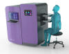
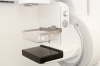
Koning Corporation
After receiving FDA clearance in November 2017 for the improvements and redesign of its Koning Breast CT (KBCT) system, the company has continued on the pathway of commercialization, adding several installations globally.