by
Christina Hwang, Contributing Reporter | August 16, 2016
A new technique that builds upon MR fingerprinting (MRF), plug-and-play MR fingerprinting (PnP-MRF), was designed by researchers from NYU Langone Medical Center, in order to create consistent quantitative data across patient populations.
MRF generally creates images from overlapping radio signals and matches the image information to databases of known tissue patterns, but the information is not always accurate.
Where MRF uses a single source of radio wave pulses to create these signals, in PnP-MRF many magnetic fields hit the tissue from all directions, so an artifact that is created in one version of the image, may not necessarily show up in another version.


Ad Statistics
Times Displayed: 46651
Times Visited: 1410 MIT labs, experts in Multi-Vendor component level repair of: MRI Coils, RF amplifiers, Gradient Amplifiers Contrast Media Injectors. System repairs, sub-assembly repairs, component level repairs, refurbish/calibrate. info@mitlabsusa.com/+1 (305) 470-8013
“Proper quantitative images can be compared directly from one patient to the next and will allow us to build a better understanding/classification of the underplaying pathology,” Martijn Cloos, Ph.D., assistant professor in the Department of Radiology at NYU Langone Medical Center, told HCB News.
“By simultaneously mapping PD, T1 and T2 we hope to increase the specificity of the diagnosis and reduce the need for invasive diagnostic procedures such as biopsies,” he said. “PnP-MRF allows one to perform rapid quantitative measurements with heterogeneous radiofrequency fields.”
According to the announcement, PnP-MRF can successfully map the distortions that occur when MR radio waves interact with tissue, and radiologists that typically erase them via calibration will no longer have this issue since PnP-MRF requires zero calibration.
“This self-calibrating nature of our method marks a clear departure from the traditional paradigm of calibration and control in MRI, and enables use of the method without fine-tuning, that is, in a ‘plug-and-play’ mode, over a broad range of experimental conditions,” Cloos said.
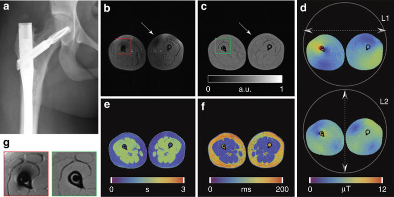
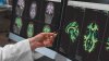
The following image compares conventional MR versus PnP-MRF for a patient with an orthopedic implant. Figure B shows an image using conventional MR, and Figures C to F are images obtained with PnP-MRF including PD, T1 and T2. Figure G shows the difference in imaging with MR and PnP-MRF.
Credit: Nature Communications, Martijn A. Cloos
Unlike traditional MR scans, where the quality of an image depends on the tissue’s exposure to the magnetic fields, the researchers said that their fingerprinting technology eliminates artifacts, leading to superior imaging with less expensive machines.
"Rather than building chambers to house extensive magnet coils that fight non-uniformities, near-future scanners, by embracing heterogeneous fields, will consist of simple tabletop magnets, or possibly even handheld MRI wands,” predicts Cloos.
The team is currently working on figuring out how to combine MRF and PET data that are acquired during a PET/MR exam.