by
Lisa Chamoff, Contributing Reporter | March 18, 2016
From the March 2016 issue of HealthCare Business News magazine
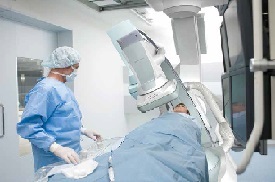
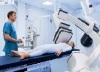
It’s not big news that over the years, cardiac catheterization labs have seen a shift, moving away from a primary focus on coronary interventions and toward less invasive structural heart procedures, such as aortic valve replacements and mitral valve repairs. Along with this change, the imaging systems for these rooms — including Xray, fluoroscopy and echocardiography — have become more sophisticated, with manufacturers promoting new features and software. For the annual focus on this special procedure room, HealthCare Business News obtained the details on what the leading manufacturers consider the best features of their cath lab equipment, and also asked clinicians which features come into play when they’re in the cath lab trenches.
“We are being asked to provide surgical-like results, if not better,” says Dr. Brijeshwar Maini, regional medical director of trans-catheter therapies for the Tenet Healthcare Corporation. “The only way you’re going to provide this is with superior imaging.”
A new view on the horizon
Multimodality imaging continues to gain ground in the hybrid suite, to guide structural heart procedures such as per-cutaneous aortic valve replacements, left atrial appendage closures (Boston Scientific received FDA approval last year for the Watchman, a left atrial appendage closure device delivered via catheter) and other image-guided, minimally invasive procedures.
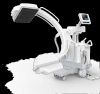
Robert Dewey, senior director of product marketing at Siemens, says that what the company calls its dynaCT Cardiac feature uses a C-arm to acquire a series of images while rotating around the region of interest, resulting in a 3-D rendering of the anatomy, which can then either be viewed side by side, or overlaid, with the fluoroscopy image. The Siemens PURE platform allows a pre-procedural CT or MR to be fused with a fluoroscopy image utilizing two reference images acquired greater than 30 degrees apart. “So if you’re introducing a valve, you have this pictorial roadmap of where to position it,” Dewey says.
Maini has used Siemens’ intracardiac echocardiography probe for procedures such as transcatheter aortic valve replacement, which he says makes the procedure much easier. In the past, when only 2-D imaging with echocardiography was available, the implantation of the valves was much more difficult, he says. “It really makes the procedure a lot easier, because now you’re not fusing those two-dimensional images in your mind and making a three-dimensional map in your brain,” Maini says. “The machine is doing that for you, which I think is a huge change. You can see your equipment, your device, in a three-dimensional plane. In my mind, I think these imaging technologies make for a superior, faster procedure. There’s still a lot of operator variability, but nonetheless, it makes for an easier, simpler experience.”