by
Gus Iversen, Editor in Chief | February 12, 2016
Courtesy:
Charité - Universitätsmedizin
Berlin
For the first time ever, researchers from Charité-Universitätsmedizin Berlin and the Max Delbrück Center for Molecular Medicine (MDC) have reportedly utilized 7 Tesla MR in a large, patient-based, cardiac study.
Cardiovascular MR (CMR) — a vital imaging procedure for precisely diagnosing myocardial diseases — is often compromised by the heart's constant movement. The researchers wanted to find out if the sharper pictures provided by the high powered magnet would allow greater insight into the condition of patients with abnormal heart muscle thickening than 3T magnets.
The physicians reported they were able to see structures on the microscopic scale, including pathological changes and small depressions in the muscles. One notable example was the ability to better assess patients with hypertrophic cardiomyopathy (HCM), a genetically determined thickening of the heart muscle.


Ad Statistics
Times Displayed: 174298
Times Visited: 3178 For those who need to move fast and expand clinical capabilities -- and would love new equipment -- the uCT 550 Advance offers a new fully configured 80-slice CT in up to 2 weeks with routine maintenance and parts and Software Upgrades for Life™ included.
Currently there are only five centers in the world that can visualize the heart with 7T MR, and their capabilities are limited to the context of research. Last year,
HCB News spoke to Siegfried Trattnig, a 7T researcher in Vienna, who said the benefits include a higher signal-to-noise ratio, a higher spectral resolution, and higher susceptibility effects to improve the spatial resolution.
Dr. Jeanette Schulz-Menger, head of the Experimental and Clinical Research Center's (ECRC) Cardiac Outpatient Department — a facility jointly run by Charité and the MDC — and head of the Working Group CMR at ECRC in cooperation with the HELIOS Klinkum Berlin-Buch, led the new research.
In a statement she said, "our aim was to test the potential of 7T MRI scanning in patients with hypertrophic cardiomyopathy, and to test whether the technology is capable of visualizing even the smallest morphological changes."
To accomplish this, they compared data obtained from patients with abnormal thickening of the heart muscle who had undergone 7T MR with 2D CINE imaging and a 3T MR scan. They also studied images of healthy volunteers taken with 7T MR.
The results, published in the latest issue of
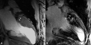
PLOS ONE, suggest that the 7T system allowed the researchers to detect "myocardial crypts" - tiny wells in muscles that had not been previously detectable.
"In seven out of 13 patients, we were able to adequately visualize minute depression in the myocardial tissue of the left ventricle," said Dr. Marcel Prothmann, the study's first author. "The technology's high spatial resolution constitutes a massive leap forward in terms of imaging quality. It allows the precise visualization of structural changes within areas of extensive thickening."
In 2015, Siemens Healthcare introduced the first 7T MR designed for clinical use, the
Magnetom Terra 7. That system is geared toward certain neurological and musculoskeletal applications.
Philips Healthcare and GE Healthcare also have 7T MR systems, but for research purposes only — Philips’ is called the Achieva 7.0T MR research system and GE’s is the Discovery MR950. In 2012, only
about 40 7T MRIs were installed worldwide — Siemens installed almost 15 in the U.S., Philips installed nine and GE installed seven in a few countries.