by
Lauren Dubinsky, Senior Reporter | November 16, 2015
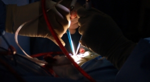
Courtesy of UA College
of Engineering
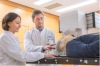
Researchers at the University of Arizona have developed a device that allows neurosurgeons to see the blood flowing inside vessels during surgery for the first time. The technology, augmented microscopy, provides surgeons with a very detailed image in real time to assist them in staying on course during surgery.
"When we started developing this technology, we thought of it like a Google map of a surgical view, providing layers of pertinent information in real time," Marek Romanowski, UA associate professor of biomedical engineering, said in a statement.
During surgery, contrast agents are administered to the patient to illuminate important diagnostic information that can prevent surgeons from severing the wrong vessel or removing healthy tissue. The technology overlaps the image a surgeon sees under a microscope with an electronically processed image using near-infrared fluorescence, which is a computer-generated imaging technology.


Ad Statistics
Times Displayed: 365069
Times Visited: 6974 Quality remanufactured Certified Centrifuges at Great prices! Fully warranted and backed by a company you can trust! Call or click for a free quote today! www.Centrifugestore.com 800-457-7576
The device is a tiny box inside of a surgical microscope that pairs electronic circuitry and optical technologies. It superimposes the fluorescence image on the real image and displays the augmented view through the microscope’s right eyepiece.
Surgeons traditionally have to look up from a surgical microscope to see the fluorescence on a display monitor. They have to switch between the real and the electronic views, and the fluorescence only shows contrast in black and white instead of anatomical structures or their spatial relationship.
The new technology displays the real and fluorescence images simultaneously and in one location. That eliminates a lot of the disruption and speculation neurosurgeons currently have to deal with.
"Our augmented technology provides diagnostic information under the microscope on demand and in color, appearing directly over tissue a surgeon is operating on — as if the tissue was painted to help direct the surgeon’s work," said Romanowski.
The most valuable use for the technology is in treating brain cancer, according to Romanowski. Brain cancer is very challenging to remove and the current surgical microscopes restrict how much of the cancer tissue surgeons can view and how accurately they can determine the tumor's borders.
If the surgeon is too aggressive, normal brain tissue may be removed and that could impair the patient’s functions, but if not enough of the tumor is removed then it may result in an immediate relapse.
Augmented microscopy may also bring benefits to the treatment of aneurysms. To treat an aneurysm, neurosurgeons seal it off from connecting vessels to prevent a rupture, but almost half of the patients with ruptured aneurysms die.
The new technology may improve their chances of survival by providing surgeons with real-time feedback on every surgical maneuver they make.