by
Lauren Dubinsky, Senior Reporter | December 17, 2014
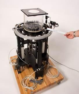
Microwave breast
tomography instrument
Courtesy of Neil Epstein
Mammography has been proven to be effective at detecting early stage tumors but it subjects patients to radiation and discomfort. A new modality without those limitations, called microwave tomography, is set to break into the industry soon.
Dartmouth College in New Hampshire is currently working on developing and refining the technology. It's still unclear exactly what role it will play in breast imaging.
During a microwave tomography exam, the breast is suspended in a liquid bath that is surrounded by 16 antennae. One at a time, each antenna illuminates the breast with a low powered microwave signal while the other antennae receive the signals that are transmitted through the breast.
That data is then used to create a 3-D model of the breast that includes the location of the normal and malignant tissue.
It has better specificity between normal and malignant tissue but mammography's spatial resolution is still superior. In order to overcome that, they're looking to pair microwave tomography with MR because of its excellent resolution.
"There is considerable interest in this technology worldwide, but the success rate of translating the concept into a clinical system is still very low," Neil R. Epstein, postdoctoral fellow at the University of Calgary in Canada, wrote to DOTmed News.
The three possible clinical breast imaging applications for it are screening, diagnosis, and therapy monitoring. Screening is the largest market but the hardest to break into because the required clinical trials are very large and expensive.
Diagnosis is a smaller market but in order for microwave tomography to be clinically accepted, it would have to be proven to perform better than biopsy. For that, Epstein is looking to pair it with MR and he noted that they have some promising results so far.
Therapy monitoring is the smallest market and Epstein believes it provides the best opportunity to break into the clinic in the shorter term. This involves imaging the breast multiple times throughout chemotherapy treatment to potentially recommend a treatment change if the tumor isn't responding.
Mammography and ultrasound have no use for that but MR and PET do. However, those modalities are invasive and expensive so the patient is usually only imaged once or twice during treatment, which isn't enough to make a treatment change recommendation.
The university is in the final stages of receiving a large reward from the National Institutes of Health to partner with a large commercial company to optimize microwave tomography for therapy monitoring and perform a multi-center clinical trial. The partner will be revealed early next week when the university officially receives the award.
Back to HCB News