From the June 2013 issue of HealthCare Business News magazine
By Kent Friedman
The medical field is always changing with new procedures, equipment and professionals. The one thing that doesn’t change, however, is the goal of providing patients with the best care possible. There are several steps that hospitals and health care centers take to ensure a high quality of service, one of which is utilizing patient simulators. For nuclear medicine, using a patient simulator (also known as a phantom) provides a unique way to test performance of nuclear medicine laboratories and interpreting physicians using clinical scenarios found in everyday practice.
The Society of Nuclear Medicine and Molecular Imaging (SNMMI) has created three nuclear medicine patient simulators — gastric emptying, PET CT chest and cardiac. The SNMMI program is the only nuclear medicine imaging patient simulator program in existence. It has been developed and operated over the past 25 years by physicians, technologists, scientists and consultants. In the last 12 years, SNMMI has worked specifically with the U.S. Department of Veterans Affairs to provide patient simulators for nuclear medicine departments. SNMMI is now making its patient simulator program available to all hospitals and facilities.
Unlike most patient simulators, nuclear medicine simulators include unknown lesions inside of them. The patient simulation exercises enhance the participants’ nuclear medicine practices by providing feedback regarding their ability to acquire, process and interpret nuclear medicine images. This is useful in comparing skills with other nuclear medicine laboratories. In addition to testing technologist and physician skills, camera systems and other technical variables can be assessed as well.
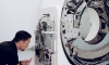
To use the patient simulator, a nuclear medicine technologist first injects an appropriate dose of radiopharmaceutical into the phantom and then scans the simulator on their camera. Once the images are acquired the lead technologist records technical data regarding the imaging session. Participating physicians view and interpret the images in the context of a clinical case scenario and are asked to answer a series of questions related to diagnostic accuracy, patient safety and appropriate use. All data is sent to the SNMMI and the exercise is graded. Remediation processes are in place for sites that do not meet the standards of image quality, or fail to pass the exercise based on other aspects of performance.