by
Kathy Mahdoubi, Senior Correspondent | May 13, 2010
"That's relatively new and a lot of research needs to be done to find out where dual energy or dual source CT plays in the overall care of patients," says Dr. Silverman. "There are many potential advantages, but dual energy CT has not yet gotten into the mainstream of clinical practice." The most benefit may be seen in cardiac and vascular imaging, but with innovation and newfound acuity come some new challenges.
"One of the major problems facing radiologists in the interpretation of images that have such high spatial resolution is finding very small mass-like lesions in many organs, including the lungs, the liver and the kidneys. Although some are clinically important, most are not. It challenges us to manage those findings in a way that is both medically appropriate and cost-effective," says Dr. Silverman.


Ad Statistics
Times Displayed: 175152
Times Visited: 3187 For those who need to move fast and expand clinical capabilities -- and would love new equipment -- the uCT 550 Advance offers a new fully configured 80-slice CT in up to 2 weeks with routine maintenance and parts and Software Upgrades for Life™ included.
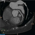
iDose overcomes limitations
(image noise) of conventional FBP
(Filtered Back Projection) reconstruction.
(Image courtesy of Philips)
Dr. Silverman is currently involved in an effort by the American College of Radiology to develop policies and recommendations for these findings. Some of the lesions may be cancerous, but Dr. Silverman says most are benign and exploration would engender many more tests at a greater cost and with added psychological strain for the patient.
In addition to the new landscape of "pseudodisease" that becomes apparent at such high resolutions, there is the sheer breadth of data acquired. There can be thousands of images to interpret, and much of the interpretation being done is not based on volumetric images.
"The truth of the matter is that the analysis of lesions and how they appear are often based on planar images, not volumetric images," states Dr. Silverman. "Volumetric, 3D images using volume-rendering or maximum intensity projection - these are useful images for looking at structures and their relationships for surgical or interventional planning, but the actual analysis of the morphology of a particular lesion is still largely dependent on its appearance on planar images. The amount of time it takes to examine the dataset is quite long and challenging. The benefits outweigh the downside, in that the image quality and the ability to use these images to diagnose are phenomenal, and that is why the use of CT in clinical practice has skyrocketed."