by
Barbara Kram, Editor | December 10, 2009
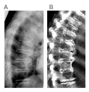
Vertebrae appear
more deformed
on VFA (right)
than radiograph
A white paper presented at the recent ASBMR annual meeting addressed a long-standing question in bone health screening: Can DXA approximate radiographs in classifying spine deformities?
It's an important question about a very prevalent, costly and debilitating health condition. (Just ask my friend, Jack who had to quit working because of degeneration of his spine.)
In the study, supported by a grant from GE, the company's new iDXA densitometer vertebral fracture assessment (VFA) was compared with conventional thoracic and lumbar spine radiographs.
Guess what?
In the hands of an experienced reader, VFA with the iDXA is comparable to using radiographs in identifying and classifying vertebral deformities in terms of cause, grade and shape. (Causes of the problem include osteoporosis, trauma, and degeneration of bone.)
For vertebral fractures, 100% agreement was found and this was higher than other DXA machines are able to achieve, perhaps because of the iDXA's double row detector design.
The study was conducted by Gabriele Armbrecht and Dieter Felsenberg at the Centre for Muscle and Bone Research, Campus Benjamin Franklin, Charite, University Medicine Berlin, Germany. Findings were presented at the American Society of Bone and Mineral Research.
It may be too late for Jack, but widespread DXA screening might help others catch and treat problems early.
Find out more at
gehealthcare.com/whybmd