
AspenOTC
Radiography market focuses on value in the COVID-19 era
November 23, 2020
by Lisa Chamoff, Contributing Reporter
Manufacturers in the radiography market continue to make workflow a priority, with new product releases featuring tube head cameras that provide for easier patient positioning as well as intuitive touchscreen displays.
They are also focusing on smaller, more price-sensitive markets, such as urgent care centers and orthopedic practices.
As always, detectors are wireless, getting lighter and able to be swapped between mobile units and fixed rooms.
Here's a look at what's new in the space.
Aspen Imaging Healthcare
Earlier this year, Aspen Imaging Healthcare released its T4 OTC, a ceiling-mounted X-ray system designed for small hospitals and orthopedic practices.
The system is designed with legs instead of a box table to allow for wheelchair accessibility. It also comes with a transparent tabletop so the technologist can ensure the detector is aligned to the tube.
The company also released its new Aspen FDR, which has the same table technology as the
AspenOTC. Both products use actuators instead of motors, which allows for "silent up and down table movement," said Rob Scorcia, vice president of marketing and business development for Aspen Imaging Healthcare.
 Canon Medical
The company recently released a set of wireless detectors called the CXDI-702C and 402C, which come with a focus on value and are compatible with both mobile and DR systems.
“Even though these are considered a value line they still include convenient hand grips,” said Lori Webb, senior manager of solutions marketing for Canon Medical.
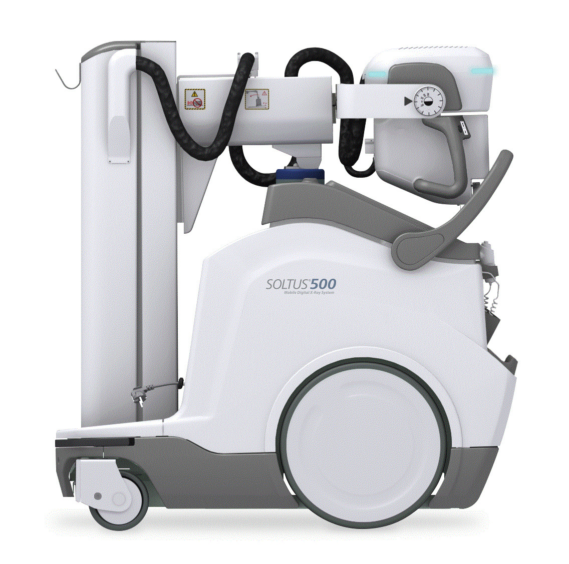
Earlier this year, Canon Medical also released its SOLTUS 500 Mobile Digital X-ray system, with a new design that is more compact and lighter than previous versions. It has a 19-inch main touchscreen display that allows for pinch-and-zoom movements and scrolling to shorten workflow, and an 8.5-inch touchscreen display on the X-ray tube head.
For infection control, the system comes with dedicated disinfecting wipe cannister holders, along with a dedicated console for charging the detector.
While the footprint is smaller and the maneuverability is more agile, there is also an anti-collision technology built into machine, with sensors that alert the end user when they’re too close to object or in a collision zone.
The system has dual-side collimator controls to shorten workflow and provide ergonomic benefits, with less bending and twisting, and also fine positioning controls at the tube head. It comes with an enhanced workflow package, allowing the technologist to toggle between the exam computer and RIS. A distributed antenna assists in connectivity challenges, providing faster image preview and less exam repeats due to a dropped signal.
 Carestream
Carestream just rolled out its new DRX Compass X-ray system, which is targeted to smaller hospitals and imaging centers, and orthopedic practices.
The system offers improved efficiency and ease of use, said Sarah Verna, worldwide marketing manager for global X-ray solutions at Carestream.
The idea behind the name Compass is that “it can take you in the direction you need to go,” Verna said.
The Compass comes with the choice of DRX Plus Detectors or Focus Detectors, and it can be used with the company’s newly released DRX-L Detector, designed specifically for long-length images of legs and spine.
The system is currently available with an overhead tube crane.
“We really responded to customers’ needs,” Verna said. “We can grow with them. You can make the room what you need for your facility.”
It also uses the most current version of Carestream’s ImageView clinical acquisition software, which recently received the Risk Management Framework Authority to Operate from the Department of Defense, a designation that necessitates fulfilling rigorous cybersecurity requirements.
On the detector side, the new DRX-L Detector is Carestream’s first single-shot, long-length imaging DR detector for leg and scoliosis exams. It is particularly ideal for pediatrics and patients with limited mobility, said Jill Hamman, worldwide marketing manager for Carestream.
The detector has a 17-inch-by-51-inch field of view and takes an exposure in one second, versus 15 seconds for traditional multiple exposure DR exams.
“It provides very high-quality images with no need for manual stitch adjustments as patient motion between shots is eliminated. This allows for a higher level of diagnostic confidence and better treatment planning,” Hamman said.
The detector features the X-Factor, which means it can be shared with other DRX Systems that do long-length imaging. Customers can purchase a portable cart to move it from room to room or mount it to a wall.
The company also released the Focus 43C (43x43 cm) wireless detector, a companion to 35C (35x43 cm) detector introduced last year. The larger size is ideal for bariatric patients as it offers a larger field of view, Hamman said. The Compass is the first X-ray system the Focus detector is integrated into. For the value tier, it provides ease of entry in moving from CR to DR.
“The introduction of our two new detectors really rounds out our detector portfolio nicely,” Hamman said.
 Del Medical
This year, Del Medical introduced its OTC18S radiographic system. One of the system’s main features is its ability to automate and improve the quality of difficult long-length leg and full spine imaging exams in a cost-effective way.
Previously, if a customer wanted automatic stitching, they needed to buy an expensive fully robotic system, said Mandy Gutierrez, Del Medical's product marketing manager.
“Typically, systems that automate stitching are only offered in the highest-end, premium, automatically positioned radiography suites,” Gutierrez said. “The OTC18S by Del Medical is a high-quality and affordable imaging solution, manufactured in America, that simplifies these complex long-length exams and is a great overall solution. This system is more affordable and offers the perfect balance between automation and manual operation.”
The system is accompanied by a newly-designed stitching barrier for patient support and immobilization. The technologist prepares the exam on the tube-side 10.4-inch touch screen. With the required image area identified, the system automatically calculates the required exposures and positions. When the technologist holds the exposure button, the OTC18s automatically moves into position, exposes, and repeats until all images are acquired and displayed fully stitched on the Delworks workstation.
Del Medical also is launching a new generation of FMT and FWFC radiographic systems. The new systems have a modern ergonomic tube head, with simplified controls and digital display of angulation and SID.
“These systems offer great value for easy to install, high-quality, American-made floor mount and floor-to-wall/floor-to-ceiling systems that provide reliable imaging in the office market,” Gutierrez said.
 Fujifilm
Fujifilm has several new products planned for RSNA, including the CALNEO Dual detector, the world’s first capture dual-layer DR detector. The detector, which the company hasn’t yet submitted for FDA clearance, allows you to generate three images from one exposure, capturing the images at different energy levels, including bone only, tissue only and traditional view.
“You can see things hidden behind the ribs,” said Rob Fabrizio, director of strategic marketing for digital radiography and women’s health at FUJIFILM Medical Systems U.S.A. Inc. “It’s a great detector for anywhere where there might be high populations of lung cancer, or for cancer treatment facilities.”
While energy subtraction has been around for long time, most energy subtraction requires two exposures.
“This is one exposure with two layers, which minimizes motion artifacts such as from the lungs and heart,” Fabrizio said.
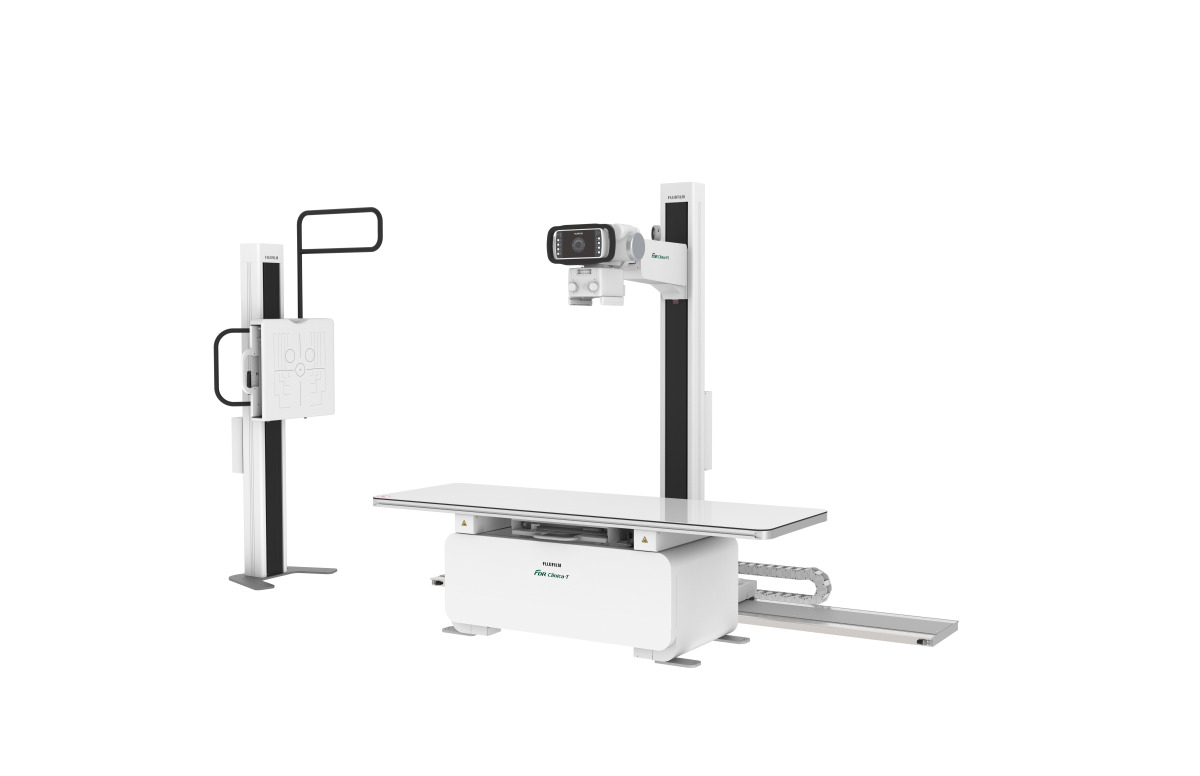
The company is also releasing three new X-ray rooms geared toward smaller, budget-minded facilities, such as outpatient centers.
As part of the Clinica family, the company is coming out with a redesigned version of its floor-mounted system, the Clinica FS. The system comes with a touch screen and auto collimation on the tube head, and X-ray generator controls built into an acquisition workstation, instead of having a secondary generator console. The system also features built-in detector connections to keep the battery charged and transfer images faster, a feature that is not typical on a low-cost floor-mounted system, Fabrizio said.
The Clinica X OTC is Fujifilm’s new overhead room, which is designed to be lower cost for smaller facilities. It also has the optional capability to perform long-length imaging at the chest stand.
“It’s a great, reliable system with the functionality you'd normally have in a hospital-grade room,” Fabrizio said.
There is also the Clinica U, a single detector U-arm system that also has a touch display on the tube head and automated exam positioning based on preselected exams.
“It brings simplified automation and consistency to the exams,” Fabrizio said.
The detector is also easily removable, which is not the case on most U-arms, according to Fabrizio.
“It’s designed for very small spaces, small budgets and exceptional versatility,” Fabrizio said. “It’s also a great backup room for hospitals.”
In the fluoroscopy space, the company is releasing a new radiography and fluoroscopy system called the Persona R&F, which is pending FDA clearance. This new multi-use R&F Room system incorporates a 17-inch-by-17-inch DR detector and brings the added ability to perform general radiography imaging.
GE Healthcare
GE Healthcare’s new Thoracic Care Suite, which is available in select CE Mark countries but not yet in the U.S., includes a suite of eight AI algorithms that scan chest X-ray findings to alert radiologists to key findings, including consolidation, which could indicate COVID-19 pneumonia. The solution is a collaboration between GE Healthcare and Lunit.
The company also launched a patient live streaming video on its fixed radiography systems. Available on Definium 656 HD and 646 HD, both ceiling-mounted systems.
“We had a customer who originally thought it might not be a big deal, but later said it dramatically changed their workflow,” said Megan Riddle-Fulton, global product marketing director for fixed X-ray at GE Healthcare.
Other new releases include advanced applications for HD systems, such as VolumeRAD, a digital X-ray tomosythesis application for use with Definium 656 HD and some older systems. A hospital in Spain used the application to distinguish mild from severe respiratory issues in suspected COVID-19 cases.
Helix 2.0 is the newest iteration of image processing for the Definium 656 HD and 646 HD, and uses AI to improve brightness and contrast to deliver consistent images regardless of external factors such as metal artifacts or exam conditions, which assists the technologist’s post-processing workflow.
GE also released its X-ray Quality Application 2.0, which aggregates data from all scanners in a radiology department and documents exam quality and number of repeats and produces a report for the entire department.
Konica Minolta
Konica Minolta’s AeroRemote Insights tool provides information on productivity, user performance and system health, including image rejection rates, dose indicators and panel drops, in order to improve workflow, accuracy and uptime. The newest release is now compatible with Konica Minolta X-ray systems driven by the company’s Ultra acquisition software, including the KDR Advanced U-Arm System.
The application is part of the trend, reinforced by the pandemic, to manage radiology departments remotely, said Guillermo Sander, director of marketing and digital radiography for Konica Minolta Healthcare Americas. It is also designed to help departments optimize personnel performance and patient satisfaction, including reducing image rejects and retakes.
“We have to make sure we do everything we need with one step,” Sander said.
The application captures data from all X-ray rooms and across multiple facilities, with the ability to view data from any computer or mobile device.
Recently, 20/20 Imaging, a division of Konica Minolta Healthcare Americas, Inc., introduced a digital tool to streamline Penning Analysis, a method to document a patient’s range of motion, for the Opal-Chiro Digital Retrofit DR solution.
Penning Analysis is considered to be the most widely accepted and utilized method for determining flexion/extension motion in a patient’s cervical spine, though it’s normally a time-consuming, manual process using extension and flexion radiographic films to measure the degree of motion between each segment of the cervical spine and occiput.
“Chiropractors have a different set of needs,” Sander said.
20/20 Imaging developed the tool in collaboration with chiropractor Daniel Lyons.
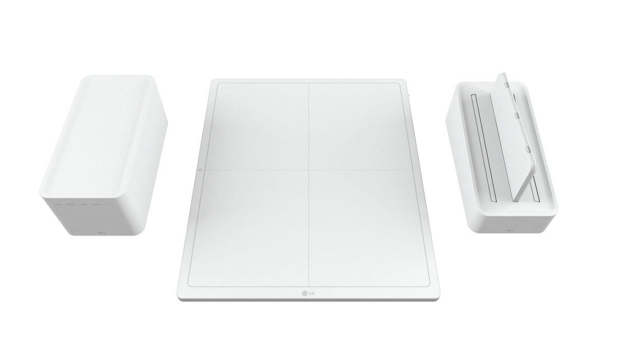
 LG Electronics
LG is new to the radiography space, but after entering into the surgical monitor space, the company began introducing its DXD flat panel detectors about two years ago.
“We saw the rad market as a good entry for LG into the medical imaging world” said Brian Whitaker, senior account manager for the ID B2B Division at LG Electronics, U.S.A. Inc. “LG’s strategy is to come in at a competitive cost, while providing better design and quality.”
The company’s latest X-ray product is a hybrid panel with fluoroscopy and general radiography capabilities, the panel will run at eight frames per second to allow for Tomographic imaging.
The panels come in 14-inch-by-17-inch wireless and 17-inch-by-17-inch wireless and tethered models. They’re made with a magnesium and carbon-fiber body, similar to the company’s TVs and refrigerators.
 Philips
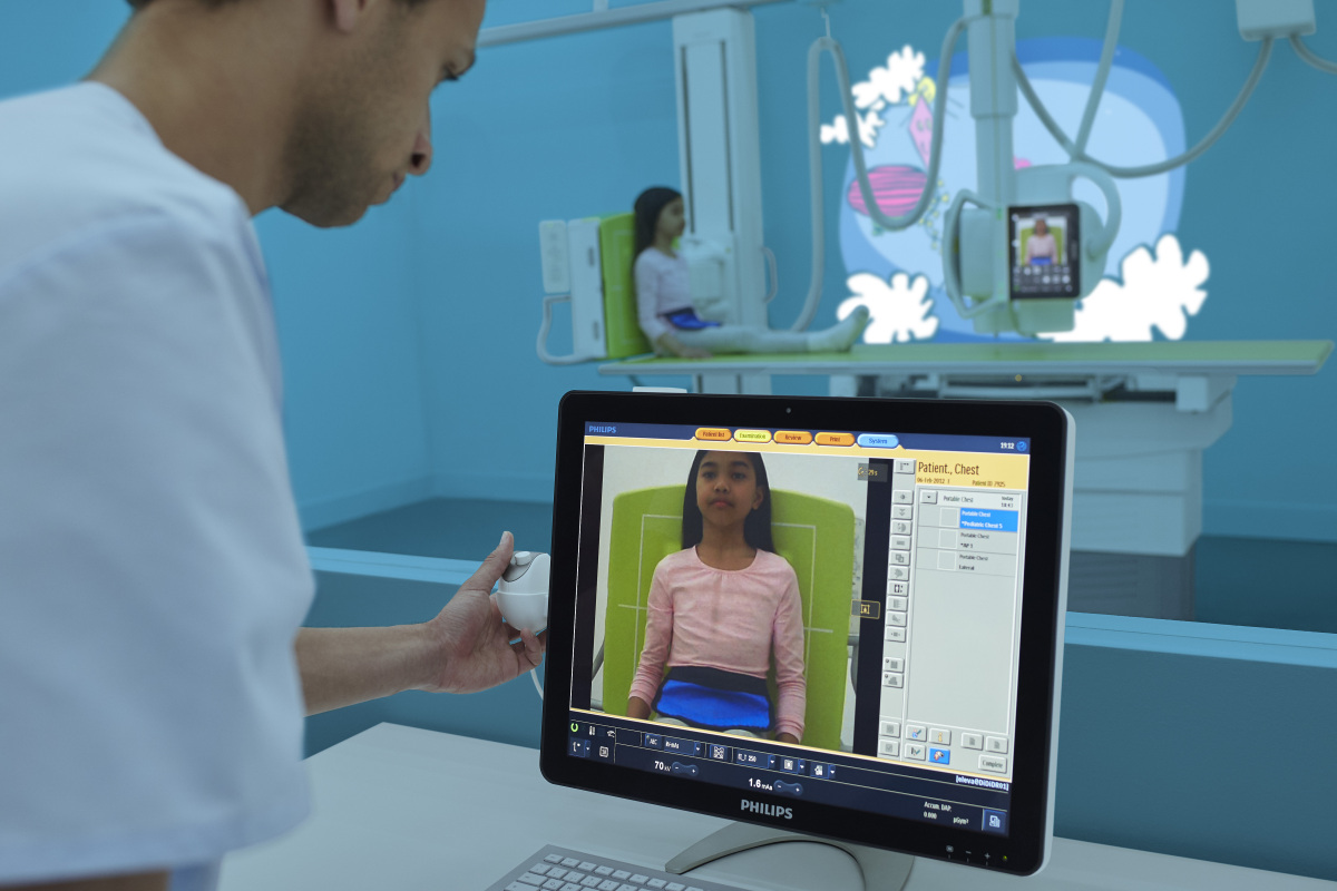
In 2018, Philips released the DigitalDiagnost C90, a ceiling-suspended radiography system that featured a tube head camera with a birds-eye view of the patient to allow for easier positioning. The company is introducing the same camera on its fluoroscopy portfolio
"We're introducing it steps-wise in our fluoro portfolio," said Daan van Manen, general manager for digital X-ray at Philips. "Our theme continues to be workflow improving patient and staff experience."
The technology is 510(k) pending and it comes with software that is the same across the entire X-ray portfolio.
"Once you're trained on one system, you can use all systems," van Manen said.
Rayence USA
At this year's RSNA virtual meeting, Rayence USA is planning to release a new line of Low-Dose TFT detectors.
The "GreenON " panels will come in a full range of sizes and offer a new standard in low-dose sensitivity.
The company will also release an upgrade to its Xmaru V1 image view software called XMaru Pro that makes it easier for users to customize their screen, quickly acquire and review images, and have some advanced processing features, said E.D. Terzi, marketing director for Rayence USA.
 Samsung
Late last year, Samsung updated its AccE portfolio of products, including the AccE GC85A ceiling-mounted DR system and AccE GM85 premium DR mobile.
The two systems were already available, launched in 2016, but were updated to have new AccE detectors, said Boris Geyzer, product manager for digital radiography at Samsung.
The new detectors have improved ergonomics, high distributed weight capacity of 882 pounds, and IP54 dust and water protection, Geyzer said.
The company is also releasing new software, including the 510(k)-pending Auto Lung Nodule Detection application, which will have its formal launch at this year’s RSNA.
Another release is its all-in-one Value Up Package, an annual update with added features and functionality to further enhance its DR systems’ clinical capabilities and workflow.
“Whenever there are new features, we incorporate that into the Value Up package,” Geyzer said. “We’re continually developing software so we can add clinically relevant features to our systems.”
Features include Voice Guide, which on the AccE GC85A allows common phrases, such as “Hold still” and “Take a deep breath” to be prerecorded so that they can easily be repeated and also programmed in a foreign language. Another feature of the Value Up software package includes the ability to add the AccE GM85’s and AccE GC85A’s S-Align angulations into the DICOM field or as part of the annotations, helping radiologists understand whether the image they are interpreting may have positioning changes that could affect the read.
At RSNA, the company is also showcasing software for real-time screen mirroring called Mirror View, for mobile X-ray used in trauma and the OR. It provides a real-time view of what’s going on on-screen so physicians and surgical staff don’t have to break the sterile field.
Siemens Healthineers
In late October, Siemens received FDA clearance for the Ysio X.pree radiography system with integrated AI. The ceiling-mounted radiography system comes equipped with myExam Companion, a user interface that automates processes and guides the technologist through the exam workflow with a 3D camera for patient positioning.
“It allows you to see depth to aid automatic thorax collimation, and will automatically raise and lower the tube head so everything is aligned,” said Martin Pesce, product manager for the X-ray products business at Siemens Healthineers North America.
The company’s new Smart Virtual Ortho application can acquire images and automatically perform stitching, eliminating the need for the technologist to set the start and end positions.
“You can place the patient on the ortho stand in front of the detector, go to the control room and adjust the collimation from the control panel,” Pesce said.
“We think this is definitely going to be the future of intelligent imaging,” Pesce said. “As a technologist myself, this is a system I would have loved to have.”
 Swissray Customer Care
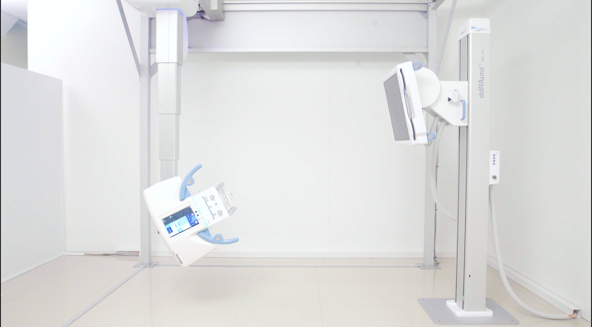
Earlier this year, Swissray Customer Care released the ddRAura OTC RT. This new system is designed to provide fast and flexible digital imaging in a busy radiography department. The system features Swissray's exclusive APS — Automated Positioning System, a motorized ceiling suspension, a smart wall stand with motorized vertical and tilt drives, and a rolling table for easy maneuvering.
The APS, coupled with the motorized drives, give the Aura OTC RT the ability to facilitate both complex and standard exams with the press of a button, according to the company. The wall stand with detector auto tracks with the central beam or moves to the off-center position programed for the specific examination and view ordered. The system will prompt the user when to remove or exchange grids.
The Aura OTC RT can be suspended from a qualified ceiling support system or it can be purchased with a preconstructed ceiling support that is floor and wall supported. This allows for the system to be installed without disturbing the finished ceiling and any HVAC, sprinkler systems or electrical components above. The preconstructed ceiling support system is called Matrix which can be customized to a room size.
The system includes a tube-mounted 9.7-inch touchscreen console for patient registration, system position, generator control and image view. The Aura OTC RT is fully configurable, with several options for generator output power, tube heat capacity, system accessories and both fixed and wireless detectors.
Thales
Since receiving CE Mark and 510(k) clearance for its ArtPix DRF System, Thales has been adding features, including the support of several sizes of dynamic and radiography portable detectors, support for angiography and stitching as well as overall improved reliability.
ArtPix DRF is an imaging platform that the company says offers high image quality and advanced clinical options.
The system includes a 17-inch-by-17-inch value detector for general radiography and two surgical panels: 12-inch-by-12-inch and 9-inch-by-9-inch format detectors. They can be plugged in to any OEM imaging system to cover all needed applications from radiography to fluoroscopy and interventional procedures.
“There was a big need for this advanced imaging system and these types of detectors to replace the existing image intensifiers platforms,” said Ilan Sivan, systems specialties and technical expertise engineer for Thales.
The company also released its ArtPix Mobile EZ2GO digital portable X-ray system embedded with Windows 10, including an upgraded security and reliability system.
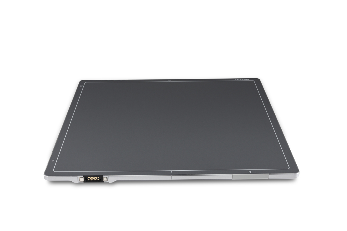
Vieworks
VIVIX-S V series are Vieworks’ newest detectors for static imaging, designed to provide a total solution for general radiography. The series has been FDA cleared and is offered in three sizes — a 25-by-30-centimeter detector called the VIVIX-S 2530VW, a 36-by-43-centimeter detector called the VIVIX-S 3643VW and a 43-by-43-centimeter detector called the VIVIX-S 4343VW.
The panels in the VIVIX-S V series are made with more advanced signal processing and superior circuitry and mechanical design to previous detectors, to provide higher image quality and better performance, still at a reasonable price, according to the company. The series also adopts an IEEE 802.11ac wireless networking standard to support faster and more stable image transmission to the image viewing software for immediate examination.
The detectors are built to be lighter and with an ergonomic design that has been recognized with a 2020 iF Product Design Award. Other new features of the series include a streamlined workflow with a variety of charging methods, longer lasting batteries that hold a charge up to 16 hours and an OLED status screen.
They are also focusing on smaller, more price-sensitive markets, such as urgent care centers and orthopedic practices.
As always, detectors are wireless, getting lighter and able to be swapped between mobile units and fixed rooms.
Here's a look at what's new in the space.
Aspen Imaging Healthcare
Earlier this year, Aspen Imaging Healthcare released its T4 OTC, a ceiling-mounted X-ray system designed for small hospitals and orthopedic practices.
The system is designed with legs instead of a box table to allow for wheelchair accessibility. It also comes with a transparent tabletop so the technologist can ensure the detector is aligned to the tube.
The company also released its new Aspen FDR, which has the same table technology as the
AspenOTC. Both products use actuators instead of motors, which allows for "silent up and down table movement," said Rob Scorcia, vice president of marketing and business development for Aspen Imaging Healthcare.
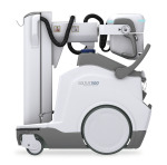
Canon Medical Systems SOLTUS 500
The company recently released a set of wireless detectors called the CXDI-702C and 402C, which come with a focus on value and are compatible with both mobile and DR systems.
“Even though these are considered a value line they still include convenient hand grips,” said Lori Webb, senior manager of solutions marketing for Canon Medical.
Earlier this year, Canon Medical also released its SOLTUS 500 Mobile Digital X-ray system, with a new design that is more compact and lighter than previous versions. It has a 19-inch main touchscreen display that allows for pinch-and-zoom movements and scrolling to shorten workflow, and an 8.5-inch touchscreen display on the X-ray tube head.
For infection control, the system comes with dedicated disinfecting wipe cannister holders, along with a dedicated console for charging the detector.
While the footprint is smaller and the maneuverability is more agile, there is also an anti-collision technology built into machine, with sensors that alert the end user when they’re too close to object or in a collision zone.
The system has dual-side collimator controls to shorten workflow and provide ergonomic benefits, with less bending and twisting, and also fine positioning controls at the tube head. It comes with an enhanced workflow package, allowing the technologist to toggle between the exam computer and RIS. A distributed antenna assists in connectivity challenges, providing faster image preview and less exam repeats due to a dropped signal.

Carestream DRX Compass
Carestream just rolled out its new DRX Compass X-ray system, which is targeted to smaller hospitals and imaging centers, and orthopedic practices.
The system offers improved efficiency and ease of use, said Sarah Verna, worldwide marketing manager for global X-ray solutions at Carestream.
The idea behind the name Compass is that “it can take you in the direction you need to go,” Verna said.
The Compass comes with the choice of DRX Plus Detectors or Focus Detectors, and it can be used with the company’s newly released DRX-L Detector, designed specifically for long-length images of legs and spine.
The system is currently available with an overhead tube crane.
“We really responded to customers’ needs,” Verna said. “We can grow with them. You can make the room what you need for your facility.”
It also uses the most current version of Carestream’s ImageView clinical acquisition software, which recently received the Risk Management Framework Authority to Operate from the Department of Defense, a designation that necessitates fulfilling rigorous cybersecurity requirements.
On the detector side, the new DRX-L Detector is Carestream’s first single-shot, long-length imaging DR detector for leg and scoliosis exams. It is particularly ideal for pediatrics and patients with limited mobility, said Jill Hamman, worldwide marketing manager for Carestream.
The detector has a 17-inch-by-51-inch field of view and takes an exposure in one second, versus 15 seconds for traditional multiple exposure DR exams.
“It provides very high-quality images with no need for manual stitch adjustments as patient motion between shots is eliminated. This allows for a higher level of diagnostic confidence and better treatment planning,” Hamman said.
The detector features the X-Factor, which means it can be shared with other DRX Systems that do long-length imaging. Customers can purchase a portable cart to move it from room to room or mount it to a wall.
The company also released the Focus 43C (43x43 cm) wireless detector, a companion to 35C (35x43 cm) detector introduced last year. The larger size is ideal for bariatric patients as it offers a larger field of view, Hamman said. The Compass is the first X-ray system the Focus detector is integrated into. For the value tier, it provides ease of entry in moving from CR to DR.
“The introduction of our two new detectors really rounds out our detector portfolio nicely,” Hamman said.

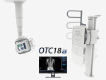
Del Medical OTC18S
This year, Del Medical introduced its OTC18S radiographic system. One of the system’s main features is its ability to automate and improve the quality of difficult long-length leg and full spine imaging exams in a cost-effective way.
Previously, if a customer wanted automatic stitching, they needed to buy an expensive fully robotic system, said Mandy Gutierrez, Del Medical's product marketing manager.
“Typically, systems that automate stitching are only offered in the highest-end, premium, automatically positioned radiography suites,” Gutierrez said. “The OTC18S by Del Medical is a high-quality and affordable imaging solution, manufactured in America, that simplifies these complex long-length exams and is a great overall solution. This system is more affordable and offers the perfect balance between automation and manual operation.”
The system is accompanied by a newly-designed stitching barrier for patient support and immobilization. The technologist prepares the exam on the tube-side 10.4-inch touch screen. With the required image area identified, the system automatically calculates the required exposures and positions. When the technologist holds the exposure button, the OTC18s automatically moves into position, exposes, and repeats until all images are acquired and displayed fully stitched on the Delworks workstation.
Del Medical also is launching a new generation of FMT and FWFC radiographic systems. The new systems have a modern ergonomic tube head, with simplified controls and digital display of angulation and SID.
“These systems offer great value for easy to install, high-quality, American-made floor mount and floor-to-wall/floor-to-ceiling systems that provide reliable imaging in the office market,” Gutierrez said.
A Clinica room from FUJIFILM
Fujifilm has several new products planned for RSNA, including the CALNEO Dual detector, the world’s first capture dual-layer DR detector. The detector, which the company hasn’t yet submitted for FDA clearance, allows you to generate three images from one exposure, capturing the images at different energy levels, including bone only, tissue only and traditional view.
“You can see things hidden behind the ribs,” said Rob Fabrizio, director of strategic marketing for digital radiography and women’s health at FUJIFILM Medical Systems U.S.A. Inc. “It’s a great detector for anywhere where there might be high populations of lung cancer, or for cancer treatment facilities.”
While energy subtraction has been around for long time, most energy subtraction requires two exposures.
“This is one exposure with two layers, which minimizes motion artifacts such as from the lungs and heart,” Fabrizio said.
The company is also releasing three new X-ray rooms geared toward smaller, budget-minded facilities, such as outpatient centers.
As part of the Clinica family, the company is coming out with a redesigned version of its floor-mounted system, the Clinica FS. The system comes with a touch screen and auto collimation on the tube head, and X-ray generator controls built into an acquisition workstation, instead of having a secondary generator console. The system also features built-in detector connections to keep the battery charged and transfer images faster, a feature that is not typical on a low-cost floor-mounted system, Fabrizio said.
The Clinica X OTC is Fujifilm’s new overhead room, which is designed to be lower cost for smaller facilities. It also has the optional capability to perform long-length imaging at the chest stand.
“It’s a great, reliable system with the functionality you'd normally have in a hospital-grade room,” Fabrizio said.
There is also the Clinica U, a single detector U-arm system that also has a touch display on the tube head and automated exam positioning based on preselected exams.
“It brings simplified automation and consistency to the exams,” Fabrizio said.
The detector is also easily removable, which is not the case on most U-arms, according to Fabrizio.
“It’s designed for very small spaces, small budgets and exceptional versatility,” Fabrizio said. “It’s also a great backup room for hospitals.”
In the fluoroscopy space, the company is releasing a new radiography and fluoroscopy system called the Persona R&F, which is pending FDA clearance. This new multi-use R&F Room system incorporates a 17-inch-by-17-inch DR detector and brings the added ability to perform general radiography imaging.
GE Healthcare
GE Healthcare’s new Thoracic Care Suite, which is available in select CE Mark countries but not yet in the U.S., includes a suite of eight AI algorithms that scan chest X-ray findings to alert radiologists to key findings, including consolidation, which could indicate COVID-19 pneumonia. The solution is a collaboration between GE Healthcare and Lunit.
The company also launched a patient live streaming video on its fixed radiography systems. Available on Definium 656 HD and 646 HD, both ceiling-mounted systems.
“We had a customer who originally thought it might not be a big deal, but later said it dramatically changed their workflow,” said Megan Riddle-Fulton, global product marketing director for fixed X-ray at GE Healthcare.
Other new releases include advanced applications for HD systems, such as VolumeRAD, a digital X-ray tomosythesis application for use with Definium 656 HD and some older systems. A hospital in Spain used the application to distinguish mild from severe respiratory issues in suspected COVID-19 cases.
Helix 2.0 is the newest iteration of image processing for the Definium 656 HD and 646 HD, and uses AI to improve brightness and contrast to deliver consistent images regardless of external factors such as metal artifacts or exam conditions, which assists the technologist’s post-processing workflow.
GE also released its X-ray Quality Application 2.0, which aggregates data from all scanners in a radiology department and documents exam quality and number of repeats and produces a report for the entire department.
Konica Minolta
Konica Minolta’s AeroRemote Insights tool provides information on productivity, user performance and system health, including image rejection rates, dose indicators and panel drops, in order to improve workflow, accuracy and uptime. The newest release is now compatible with Konica Minolta X-ray systems driven by the company’s Ultra acquisition software, including the KDR Advanced U-Arm System.
The application is part of the trend, reinforced by the pandemic, to manage radiology departments remotely, said Guillermo Sander, director of marketing and digital radiography for Konica Minolta Healthcare Americas. It is also designed to help departments optimize personnel performance and patient satisfaction, including reducing image rejects and retakes.
“We have to make sure we do everything we need with one step,” Sander said.
The application captures data from all X-ray rooms and across multiple facilities, with the ability to view data from any computer or mobile device.
Recently, 20/20 Imaging, a division of Konica Minolta Healthcare Americas, Inc., introduced a digital tool to streamline Penning Analysis, a method to document a patient’s range of motion, for the Opal-Chiro Digital Retrofit DR solution.
Penning Analysis is considered to be the most widely accepted and utilized method for determining flexion/extension motion in a patient’s cervical spine, though it’s normally a time-consuming, manual process using extension and flexion radiographic films to measure the degree of motion between each segment of the cervical spine and occiput.
“Chiropractors have a different set of needs,” Sander said.
20/20 Imaging developed the tool in collaboration with chiropractor Daniel Lyons.
LG DXD
LG is new to the radiography space, but after entering into the surgical monitor space, the company began introducing its DXD flat panel detectors about two years ago.
“We saw the rad market as a good entry for LG into the medical imaging world” said Brian Whitaker, senior account manager for the ID B2B Division at LG Electronics, U.S.A. Inc. “LG’s strategy is to come in at a competitive cost, while providing better design and quality.”
The company’s latest X-ray product is a hybrid panel with fluoroscopy and general radiography capabilities, the panel will run at eight frames per second to allow for Tomographic imaging.
The panels come in 14-inch-by-17-inch wireless and 17-inch-by-17-inch wireless and tethered models. They’re made with a magnesium and carbon-fiber body, similar to the company’s TVs and refrigerators.
Philips DigitalDiagnostC90
In 2018, Philips released the DigitalDiagnost C90, a ceiling-suspended radiography system that featured a tube head camera with a birds-eye view of the patient to allow for easier positioning. The company is introducing the same camera on its fluoroscopy portfolio
"We're introducing it steps-wise in our fluoro portfolio," said Daan van Manen, general manager for digital X-ray at Philips. "Our theme continues to be workflow improving patient and staff experience."
The technology is 510(k) pending and it comes with software that is the same across the entire X-ray portfolio.
"Once you're trained on one system, you can use all systems," van Manen said.
Rayence USA
At this year's RSNA virtual meeting, Rayence USA is planning to release a new line of Low-Dose TFT detectors.
The "GreenON " panels will come in a full range of sizes and offer a new standard in low-dose sensitivity.
The company will also release an upgrade to its Xmaru V1 image view software called XMaru Pro that makes it easier for users to customize their screen, quickly acquire and review images, and have some advanced processing features, said E.D. Terzi, marketing director for Rayence USA.
Samsung AccE
Late last year, Samsung updated its AccE portfolio of products, including the AccE GC85A ceiling-mounted DR system and AccE GM85 premium DR mobile.
The two systems were already available, launched in 2016, but were updated to have new AccE detectors, said Boris Geyzer, product manager for digital radiography at Samsung.
The new detectors have improved ergonomics, high distributed weight capacity of 882 pounds, and IP54 dust and water protection, Geyzer said.
The company is also releasing new software, including the 510(k)-pending Auto Lung Nodule Detection application, which will have its formal launch at this year’s RSNA.
Another release is its all-in-one Value Up Package, an annual update with added features and functionality to further enhance its DR systems’ clinical capabilities and workflow.
“Whenever there are new features, we incorporate that into the Value Up package,” Geyzer said. “We’re continually developing software so we can add clinically relevant features to our systems.”
Features include Voice Guide, which on the AccE GC85A allows common phrases, such as “Hold still” and “Take a deep breath” to be prerecorded so that they can easily be repeated and also programmed in a foreign language. Another feature of the Value Up software package includes the ability to add the AccE GM85’s and AccE GC85A’s S-Align angulations into the DICOM field or as part of the annotations, helping radiologists understand whether the image they are interpreting may have positioning changes that could affect the read.
At RSNA, the company is also showcasing software for real-time screen mirroring called Mirror View, for mobile X-ray used in trauma and the OR. It provides a real-time view of what’s going on on-screen so physicians and surgical staff don’t have to break the sterile field.
Siemens Healthineers
In late October, Siemens received FDA clearance for the Ysio X.pree radiography system with integrated AI. The ceiling-mounted radiography system comes equipped with myExam Companion, a user interface that automates processes and guides the technologist through the exam workflow with a 3D camera for patient positioning.
“It allows you to see depth to aid automatic thorax collimation, and will automatically raise and lower the tube head so everything is aligned,” said Martin Pesce, product manager for the X-ray products business at Siemens Healthineers North America.
The company’s new Smart Virtual Ortho application can acquire images and automatically perform stitching, eliminating the need for the technologist to set the start and end positions.
“You can place the patient on the ortho stand in front of the detector, go to the control room and adjust the collimation from the control panel,” Pesce said.
“We think this is definitely going to be the future of intelligent imaging,” Pesce said. “As a technologist myself, this is a system I would have loved to have.”
Earlier this year, Swissray Customer Care released the ddRAura OTC RT. This new system is designed to provide fast and flexible digital imaging in a busy radiography department. The system features Swissray's exclusive APS — Automated Positioning System, a motorized ceiling suspension, a smart wall stand with motorized vertical and tilt drives, and a rolling table for easy maneuvering.
The APS, coupled with the motorized drives, give the Aura OTC RT the ability to facilitate both complex and standard exams with the press of a button, according to the company. The wall stand with detector auto tracks with the central beam or moves to the off-center position programed for the specific examination and view ordered. The system will prompt the user when to remove or exchange grids.
The Aura OTC RT can be suspended from a qualified ceiling support system or it can be purchased with a preconstructed ceiling support that is floor and wall supported. This allows for the system to be installed without disturbing the finished ceiling and any HVAC, sprinkler systems or electrical components above. The preconstructed ceiling support system is called Matrix which can be customized to a room size.
The system includes a tube-mounted 9.7-inch touchscreen console for patient registration, system position, generator control and image view. The Aura OTC RT is fully configurable, with several options for generator output power, tube heat capacity, system accessories and both fixed and wireless detectors.
Thales
Since receiving CE Mark and 510(k) clearance for its ArtPix DRF System, Thales has been adding features, including the support of several sizes of dynamic and radiography portable detectors, support for angiography and stitching as well as overall improved reliability.
ArtPix DRF is an imaging platform that the company says offers high image quality and advanced clinical options.
The system includes a 17-inch-by-17-inch value detector for general radiography and two surgical panels: 12-inch-by-12-inch and 9-inch-by-9-inch format detectors. They can be plugged in to any OEM imaging system to cover all needed applications from radiography to fluoroscopy and interventional procedures.
“There was a big need for this advanced imaging system and these types of detectors to replace the existing image intensifiers platforms,” said Ilan Sivan, systems specialties and technical expertise engineer for Thales.
The company also released its ArtPix Mobile EZ2GO digital portable X-ray system embedded with Windows 10, including an upgraded security and reliability system.
Vieworks
VIVIX-S V series are Vieworks’ newest detectors for static imaging, designed to provide a total solution for general radiography. The series has been FDA cleared and is offered in three sizes — a 25-by-30-centimeter detector called the VIVIX-S 2530VW, a 36-by-43-centimeter detector called the VIVIX-S 3643VW and a 43-by-43-centimeter detector called the VIVIX-S 4343VW.
The panels in the VIVIX-S V series are made with more advanced signal processing and superior circuitry and mechanical design to previous detectors, to provide higher image quality and better performance, still at a reasonable price, according to the company. The series also adopts an IEEE 802.11ac wireless networking standard to support faster and more stable image transmission to the image viewing software for immediate examination.
The detectors are built to be lighter and with an ergonomic design that has been recognized with a 2020 iF Product Design Award. Other new features of the series include a streamlined workflow with a variety of charging methods, longer lasting batteries that hold a charge up to 16 hours and an OLED status screen.