Konica Minolta and Shimadzu are
unveiling DDR on the RADSpeed Pro
X-ray system at AHRA
unveiling DDR on the RADSpeed Pro
X-ray system at AHRA
Konica Minolta and Shimadzu bring Dynamic Digital Radiography to RADspeed Pro at AHRA
July 22, 2019
by John R. Fischer, Senior Reporter
Konica Minolta Healthcare Americas is accelerating the sale and distribution of its Dynamic Digital Radiography (DDR) solution for X-ray in the U.S. healthcare market with the help of long-time partner Shimadzu Medical Systems.
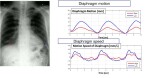
Utilizing Konica Minolta’s new advanced image processing capabilities with Shimadzu’s RADspeed Pro radiographic imaging systems, the technology provides users with a "movie" that shows the motion of structures such as bones and lungs. It will make its official debut on RADspeed Pro this week at the 45th Annual Meeting of the Association for Medical Imaging Management (AHRA).
“The only way to get the equivalent of a ‘movie’ is with extremely high dose and varied workflow, which is inconvenient for the radiologist and means a much larger procedure for the patient. Using the unique Konica Minolta technology and smart X-ray capabilities for image processing, we can produce at almost the same dose as a standard X-ray a moving image,” David Widmann, president and CEO of Konica Minolta Healthcare Americas, told HCB News. “The radiologist, pulmonologist and orthopedist can actually see the dynamics of the structure they’re looking at. It revolutionizes the idea of X-ray, this 124-year-old modality.”
Identifying movement in X-ray often requires users to take two or more images and compare the location of structures. DDR enables clinicians to analyze and quantify the dynamic interaction of anatomical structures with physiological changes over time, enhancing diagnostic capability and efficacy.
The solution at the moment is primarily meant for assessing pulmonary function and mechanics, and underwent a number of clinical studies at the Icahn School of Medicine at Mount Sinai in New York. Researchers there reported greater contextual understanding of dyspnea and other pathophysiologic abnormalities with the solution, as well as an earlier and more thorough understanding of the etiology of dyspnea.
The Institutional Review Board (IRB) approved study consisted of 43 patients. Of these, 16 were diagnosed with diaphragm abnormalities, of which six were minimally or not at all visible on a conventional chest X-ray. The ten that were visible on the conventional chest X-ray were better defined on DDR, which also identified two cases of chronic obstructive pulmonary disease (COPD) that were not detected on the chest X-ray. Prior studies from Mount Sinai show that DDR may be clinically relevant for assessing the severity of COPD in acute settings, and for patients unable to perform pulmonary further testing.
“X-ray exams are primary imaging exams and account for 70 percent of all imaging procedures today. They are done before you go for a CT or MR. Having more insight in that particular exam provides for better patient diagnosis, less time in the healthcare cycle and more precise diagnosis and treatment plans for the individual,” said Widmann. “That’s really our precision aim with this technology.”
He notes that plans are in place to apply the technology for other applications, systems and specialties, with a second application for orthopedics expected in the fall.
“That will bring us squarely in the middle of not only a hospital setting but also every orthopedic and pain clinic across the U.S. You can see joints moving and determine conflict between bone and tendons,” he said. “We think that right now DDR can be an application that can be applied to not only many different systems but also many different clinical applications.”
Konica Minolta and Shimadzu will introduce DDR on the RADSpeed Pro this week at the 45th Annual Meeting of the Association for Medical Imaging Management (AHRA).