
Daniel Merton
OB/GYN ultrasound: is the field ready to adopt new technology in the market?
July 08, 2019
by Lauren Dubinsky, Senior Reporter
Automated data acquisition tools for ultrasound can automatically gather measurements, potentially reducing exam times and allowing clinicians to spend more time interfacing with their patients. But are these tools actually being used in the clinical setting?
“If you talk to professionals in different maternal fetal medicine laboratories, you're going to find that they are not universally using [these] tools,” said Daniel A. Merton, principal project officer at ECRI Institute. “As beneficial as they might sound, unfortunately, people are creatures of habit, and it’s sometimes challenging to break those habits.”
He has been conducting research in the ultrasound field for over 30 years and has seen this trend quite often. Historically, technologies with potential benefits like these automation tools, sometimes don’t make their way into routine clinical utilization because the field doesn’t believe the benefits outweigh the downsides.
 “The downside to new technologies is that they require a different way of performing examinations,” said Merton. “If you've been doing the exam the old-fashioned, manual way for 20 years, chances are that you're going to continue to do it that way.” For instance, if a sonographer tries a new measurement tool a few times and finds that it doesn't put the cursors in the right location, they’re most likely not going to continue to use it.
However, the technologies that are clinically proved to be useful will eventually be adopted.
“I'm convinced that this is the way that's going to go with automation in OB ultrasound,” said Merton. “It may not happen overnight. These things generally don't happen overnight, but within five to 10 years it's going to be commonplace.”
A provider must have a lot of trust in a given system in order to allow it that much control over exams. Semi-automated technology is one way to create a better balance so that the sonographer and the technology can better understand each other.
“I think a lot of sonographers are very suspicious of anything that does it for them because it’s not the easiest thing to do sometimes,” said Tina Hodgson, senior manager of solutions marketing for ultrasound at Canon Medical Systems USA, which offers semi-automated measurement capabilities for fetal biometry, but chooses not to offer completely automated tools.
Philips Healthcare, on the other hand, offers its aBiometry Assist application, which uses anatomical intelligence of fetal anatomy to automatically pre-place measurement cursors on selected structures that clinicians can accept or edit. Despite the automated nature of the technology, Philips is seeing enthusiastic adoption of these automation tools.
Companies excited about potential of AI
One area stirring up a lot of excitement is the use of artificial intelligence in OB/GYN ultrasound and the significant role it could play in the coming years for both new and existing users.
“For high-volume practices, AI will aid in automating exams to rule out anomalies,” said Jeff Cohen, general imaging and women’s healthcare ultrasound business leader at Philips. “This will enable clinicians to spend more time with their patients and on their more complex cases.”
It could also play a role in providing the initial analysis of the severity of a fetal anomaly, and help make OB/GYN ultrasound more accessible for less skilled users because of its potential to automate exams.
“This will play an ever-increasing role in developing and emerging markets where governments are investing in expanding access to healthcare,” said Cohen. “The challenge beyond equipment and solutions is skilled users.”
GE now offers its SonoCNS technology on its Voluson ultrasound systems to help properly align and display recommended views and measurements of the fetal brain. This is part of GE’s Edison Platform, which is designed to integrate and assimilate data from disparate sources, apply advanced analytics and AI to the data, and generate insights to support clinical, financial and operational decision-making.
 “Advancements in AI and automation simplify the exam process for the user, while gaining result consistency and reproducibility,” said Roland Rott, general manager of GE’s women’s health ultrasound. “Also, clinicians are looking to image earlier so that anomalies can be detected sooner, which can enhance patient management and outcomes.”
Reducing work-related injuries for sonographers
Work related musculoskeletal disorders (WRMSDs) affect up to 90 percent of sonographers and other users of diagnostic medical sonography, according to the Society of Diagnostic Medical Sonography (SDMS). They can take a significant personal toll on the affected because they might no longer be able to work or perform simple, daily tasks.
Merton explained that sonographers are required to put pressure down with their arm extended away from their body, which causes muscular stress. This could result in an injury to the wrist, shoulder, neck, elbow or a combination thereof.
“In terms of OB scanning, as the patient's abdomen gets larger, and if you're a smaller person performing the exam, you may have your arm at a 90-degree angle out from your body while trying to push down,” said Merton. “That's very difficult to do and very stressful on the body.”
Canon’s intelligent Dynamic Micro-Slice (iDMS) technology takes aim at reducing these injuries by allowing for extra penetration while being able to scan at a higher frequency to get better resolution and reduce the noise in the image.
“When you get third trimester patients, you’re sometimes struggling to get a good view of those babies if you have a large patient,” said Hodgson. “We found that with our new IDMS technology, that made an enormous difference.”
 A major factor contributing to WRMSDs is the increase in exams that sonographers are being asked to perform throughout a given shift. They used to scan six to eight patients in an eight-hour day, but now they might be scanning up to 12 or more patients per day.
In addition, there is not much downtime between the exams for the sonographer to rest, because of digital imaging. With film imaging, the sonographers would need to run their films and reload their cassettes, which gave them a 10- to 15-minute break.
The Industry Standards for the Prevention of Work-Related Musculoskeletal Disorders in Sonography was developed through a consensus conference hosted by the SDMS in 2016, which ECRI helped revise in 2017. The standards, which are available online, state that working posture and the ergonomics of the work environment are crucial to preventing an injury or managing the progression of symptoms.
Benefits of early and late pregnancy ultrasounds
Most women get an ultrasound in their second trimester, but a new study conducted by the University of Cambridge and the University of East Anglia found that late pregnancy ultrasounds may also result in additional benefits and cost savings.
Currently, physicians and midwives feel the mother’s stomach to determine the position of the baby to check for breech presentation, but the sensitivity and accuracy of this method depends on the practitioner.
“It is well recognized that a significant proportion of cases of breech presentation are missed using this method,” said Gordon Smith, professor of obstetrics and gynaecology at the University of Cambridge and chief investigator of the study.
 Smith and his research team performed screening ultrasound exams at 36 weeks gestation in 3,879 women having first pregnancies in England. They diagnosed 179 women with breech presentation, but in 55 percent of the cases there was no prior suspicion that the baby was in the breech position.
The team also considered the cost of additional ultrasound scans and found that if £12.90 was charged per scan, it could be cost-saving for the National Health Service.
Smith explained that the cost of ultrasound significantly varies based on the country and the complexity of the scan. A complex ultrasound scan cannot be performed for £12.90, but it might be possible for midwives to perform a presentation scan using a point-of-care ultrasound.
“I think that it is likely that point-of-care ultrasound will replace clinical examination to detect breech presentation,” he added. “The time horizon will depend on the technology. If there was a simple probe that could Bluetooth connect to a smartphone and which did not carry huge costs, I could see this becoming quite widespread within five years or less.”
 Dr. Stephen T. Chasen, maternal/fetal medicine specialist at Weill Cornell Medicine and NewYork-Presbyterian, believes that routine ultrasound could avoid undiagnosed breech presentation.
“Most obstetricians have ultrasound in their office,” he said. “Unlike detecting fetal abnormalities, determining fetal presentation with office ultrasound is well within the training and expertise of general OB-GYNs, and implementing such a protocol would generally not require outside referral.”
Chasen is also a supportive of performing ultrasound scans in the first trimester to identify major abnormalities early in pregnancy. He explained that many structures can be diagnosed before 14 weeks.
He warned against the temptation to solely follow guidelines from professional organizations and payors. If these exams are simply a checklist from the insurer, then abnormalities in the face, heart, extremities and genitalia can be missed.
“Guidelines from professional societies tend to be relatively simple and probably don't set the bar too high in terms of quality,” said Chasen. “They recognize the variation in practice settings and expertise and probably don't want to expose their members to medico-legal liability. The temptation to adhere to the guidelines of payers largely relates to maximizing revenue.”
Ultrasound-guided epidural injections
Charlottesville-based Rivanna Medical introduced an ultrasound device to the market in 2016 called Accuro, which leverages ultrasound technology to guide epidural injections.
Traditionally, physicians used a technique that involved counting down the number of bones on the patient’s spine to identify the space where the anesthesia should be administered. Once the appropriate level is identified, the physician advances the needle until they reach the correct depth to numb the roots of the nerves for the legs and pelvis.
“Doing the procedure using this technique is a little like throwing a dart across the room with your eyes closed,” said ECRI’s Merton.
Conventional ultrasound systems have been used to guide these injections, but it requires the user to have a lot of skill and experience. The Accuro device provides a 3D display of the location of the bones, the intervertebral space, and indicates the depth that the anesthetic needs to be administered.
 “Over the past two decades, the field of anesthesia has migrated toward an image guidance standard of care for needle placement procedures such as vascular access and peripheral nerve blockades,” said Will Mauldin, CEO of Rivanna Medical. “This procedure remains blind despite an abundance of academic literature, including meta-analysis, that demonstrates improved success rates, reduced needle sticks, and improved safety when ultrasound is used to guide the neuraxial placement.”
Anesthesia providers still prefer the blind approach over image guidance because ultrasound isn’t great at visualizing bone, the technology is not always accessible, and there is an extreme learning curve for spinal ultrasound, he added.
Accuro was developed to fill that gap, but as with any new technology the anesthesia community has approached it with caution. However, adoption has rapidly increased after a University of Virginia trial demonstrated improved success rates, patient satisfaction, and needle insertion times compared to the standard approach.
“There is no question that Accuro is driving neuraxial placements toward the same image-guided standard of care already in place for vascular access and peripheral nerve blockades,” said Mauldin. “We expect that major anesthesia organizations will begin to integrate image guidance into official clinical guidelines.”
“If you talk to professionals in different maternal fetal medicine laboratories, you're going to find that they are not universally using [these] tools,” said Daniel A. Merton, principal project officer at ECRI Institute. “As beneficial as they might sound, unfortunately, people are creatures of habit, and it’s sometimes challenging to break those habits.”
He has been conducting research in the ultrasound field for over 30 years and has seen this trend quite often. Historically, technologies with potential benefits like these automation tools, sometimes don’t make their way into routine clinical utilization because the field doesn’t believe the benefits outweigh the downsides.

Daniel Merton
However, the technologies that are clinically proved to be useful will eventually be adopted.
“I'm convinced that this is the way that's going to go with automation in OB ultrasound,” said Merton. “It may not happen overnight. These things generally don't happen overnight, but within five to 10 years it's going to be commonplace.”
A provider must have a lot of trust in a given system in order to allow it that much control over exams. Semi-automated technology is one way to create a better balance so that the sonographer and the technology can better understand each other.
“I think a lot of sonographers are very suspicious of anything that does it for them because it’s not the easiest thing to do sometimes,” said Tina Hodgson, senior manager of solutions marketing for ultrasound at Canon Medical Systems USA, which offers semi-automated measurement capabilities for fetal biometry, but chooses not to offer completely automated tools.
Philips Healthcare, on the other hand, offers its aBiometry Assist application, which uses anatomical intelligence of fetal anatomy to automatically pre-place measurement cursors on selected structures that clinicians can accept or edit. Despite the automated nature of the technology, Philips is seeing enthusiastic adoption of these automation tools.
Companies excited about potential of AI
One area stirring up a lot of excitement is the use of artificial intelligence in OB/GYN ultrasound and the significant role it could play in the coming years for both new and existing users.
“For high-volume practices, AI will aid in automating exams to rule out anomalies,” said Jeff Cohen, general imaging and women’s healthcare ultrasound business leader at Philips. “This will enable clinicians to spend more time with their patients and on their more complex cases.”
It could also play a role in providing the initial analysis of the severity of a fetal anomaly, and help make OB/GYN ultrasound more accessible for less skilled users because of its potential to automate exams.
“This will play an ever-increasing role in developing and emerging markets where governments are investing in expanding access to healthcare,” said Cohen. “The challenge beyond equipment and solutions is skilled users.”
GE now offers its SonoCNS technology on its Voluson ultrasound systems to help properly align and display recommended views and measurements of the fetal brain. This is part of GE’s Edison Platform, which is designed to integrate and assimilate data from disparate sources, apply advanced analytics and AI to the data, and generate insights to support clinical, financial and operational decision-making.

Roland Rott
Reducing work-related injuries for sonographers
Work related musculoskeletal disorders (WRMSDs) affect up to 90 percent of sonographers and other users of diagnostic medical sonography, according to the Society of Diagnostic Medical Sonography (SDMS). They can take a significant personal toll on the affected because they might no longer be able to work or perform simple, daily tasks.
Merton explained that sonographers are required to put pressure down with their arm extended away from their body, which causes muscular stress. This could result in an injury to the wrist, shoulder, neck, elbow or a combination thereof.
“In terms of OB scanning, as the patient's abdomen gets larger, and if you're a smaller person performing the exam, you may have your arm at a 90-degree angle out from your body while trying to push down,” said Merton. “That's very difficult to do and very stressful on the body.”
Canon’s intelligent Dynamic Micro-Slice (iDMS) technology takes aim at reducing these injuries by allowing for extra penetration while being able to scan at a higher frequency to get better resolution and reduce the noise in the image.
“When you get third trimester patients, you’re sometimes struggling to get a good view of those babies if you have a large patient,” said Hodgson. “We found that with our new IDMS technology, that made an enormous difference.”

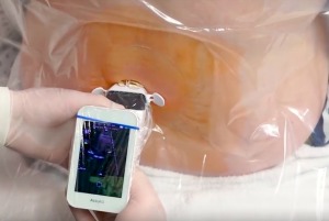
A physician using Rivanna Medical's Accuro device.
In addition, there is not much downtime between the exams for the sonographer to rest, because of digital imaging. With film imaging, the sonographers would need to run their films and reload their cassettes, which gave them a 10- to 15-minute break.
The Industry Standards for the Prevention of Work-Related Musculoskeletal Disorders in Sonography was developed through a consensus conference hosted by the SDMS in 2016, which ECRI helped revise in 2017. The standards, which are available online, state that working posture and the ergonomics of the work environment are crucial to preventing an injury or managing the progression of symptoms.
Benefits of early and late pregnancy ultrasounds
Most women get an ultrasound in their second trimester, but a new study conducted by the University of Cambridge and the University of East Anglia found that late pregnancy ultrasounds may also result in additional benefits and cost savings.
Currently, physicians and midwives feel the mother’s stomach to determine the position of the baby to check for breech presentation, but the sensitivity and accuracy of this method depends on the practitioner.
“It is well recognized that a significant proportion of cases of breech presentation are missed using this method,” said Gordon Smith, professor of obstetrics and gynaecology at the University of Cambridge and chief investigator of the study.

Gordon Smith
The team also considered the cost of additional ultrasound scans and found that if £12.90 was charged per scan, it could be cost-saving for the National Health Service.
Smith explained that the cost of ultrasound significantly varies based on the country and the complexity of the scan. A complex ultrasound scan cannot be performed for £12.90, but it might be possible for midwives to perform a presentation scan using a point-of-care ultrasound.
“I think that it is likely that point-of-care ultrasound will replace clinical examination to detect breech presentation,” he added. “The time horizon will depend on the technology. If there was a simple probe that could Bluetooth connect to a smartphone and which did not carry huge costs, I could see this becoming quite widespread within five years or less.”

Dr. Stephen Chasen
“Most obstetricians have ultrasound in their office,” he said. “Unlike detecting fetal abnormalities, determining fetal presentation with office ultrasound is well within the training and expertise of general OB-GYNs, and implementing such a protocol would generally not require outside referral.”
Chasen is also a supportive of performing ultrasound scans in the first trimester to identify major abnormalities early in pregnancy. He explained that many structures can be diagnosed before 14 weeks.
He warned against the temptation to solely follow guidelines from professional organizations and payors. If these exams are simply a checklist from the insurer, then abnormalities in the face, heart, extremities and genitalia can be missed.
“Guidelines from professional societies tend to be relatively simple and probably don't set the bar too high in terms of quality,” said Chasen. “They recognize the variation in practice settings and expertise and probably don't want to expose their members to medico-legal liability. The temptation to adhere to the guidelines of payers largely relates to maximizing revenue.”
Ultrasound-guided epidural injections
Charlottesville-based Rivanna Medical introduced an ultrasound device to the market in 2016 called Accuro, which leverages ultrasound technology to guide epidural injections.
Traditionally, physicians used a technique that involved counting down the number of bones on the patient’s spine to identify the space where the anesthesia should be administered. Once the appropriate level is identified, the physician advances the needle until they reach the correct depth to numb the roots of the nerves for the legs and pelvis.
“Doing the procedure using this technique is a little like throwing a dart across the room with your eyes closed,” said ECRI’s Merton.
Conventional ultrasound systems have been used to guide these injections, but it requires the user to have a lot of skill and experience. The Accuro device provides a 3D display of the location of the bones, the intervertebral space, and indicates the depth that the anesthetic needs to be administered.

Will Mauldin
Anesthesia providers still prefer the blind approach over image guidance because ultrasound isn’t great at visualizing bone, the technology is not always accessible, and there is an extreme learning curve for spinal ultrasound, he added.
Accuro was developed to fill that gap, but as with any new technology the anesthesia community has approached it with caution. However, adoption has rapidly increased after a University of Virginia trial demonstrated improved success rates, patient satisfaction, and needle insertion times compared to the standard approach.
“There is no question that Accuro is driving neuraxial placements toward the same image-guided standard of care already in place for vascular access and peripheral nerve blockades,” said Mauldin. “We expect that major anesthesia organizations will begin to integrate image guidance into official clinical guidelines.”