
Ramsey D. Badawi
The whole body PET scanner is getting closer to becoming a reality
June 20, 2017
Ramsey D. Badawi, professor and molecular imaging chair in radiology at the University of California, Davis, and chief of nuclear medicine, and Simon R. Cherry, Ph.D., a distinguished professor of biomedical engineering and radiology at UC, Davis, are two men with a single vision: to build a PET scanner large enough to image an entire human in a single scan. The mission to create this “whole body” PET scanner became their passion – and now they’re on the verge of seeing that vision come true. HealthCare Business News interviewed the two men so they could detail how they are meeting this challenge.
HCB News: How did this whole body PET project initially get started?
Badawi & Cherry: In the fall of 2005, we were discussing future projects that we could collaborate on. I mentioned that I had done some simulations of long PET scanners in the past and that we should try to build one, perhaps 60 cm long or so. Simon replied that if we were to do this, we should go the whole hog and build a two-meter one that would cover the entire body! We both got very excited about this idea and decided to pursue it. We launched the project in 2006, when Simon described the idea at the Henry Wagner Jr. lecture at the SNMMI annual meeting. It took us 10 years from the time of the initial idea to finally obtaining funding from the NIH to build the first prototype.
HCB News: What are the perceived benefits from whole body PET compared to conventional PET imaging?
B & C: There are two main features that lead to a range of benefits: for total body applications, one can collect ~40x as much signal as with a conventional scanner (or about 5x more signal if one is only interested in a single organ) and one can scan the entire body simultaneously.
Due to the increased sensitivity to signal, one can either:
 * Scan more gently. We can use the sensitivity to use less radioactive material. This is very important for pediatric patients, or for doing scientific studies where we want to scan the same patient many times to look for changes. Examples would be learning more about the biology and cures for arthritis, or diabetes or obesity, where the diseases have time courses of many years.
In addition, one can scan everything. The ability to scan the entire body at the same time allows us to make movies showing, for example, how drugs move around inside the body. This allows us to see to what degree new drugs hit their target, and to what degree they hit non-target organs, causing side-effects. We can also learn about interactions and signaling between different organ systems. For example, the brain and the gut. This could prove to be very important for Parkinson's disease, for Alzheimer's disease, and for obesity, among others.
HCB News: What hurdles stand in the way of completing this project? Do you have a completion date in mind?
B & C: This will be the biggest, most complex PET scanner for human use yet built. The scanner has over 560,000 detector elements and almost 54,000 light sensors and channels of electronics. There will always be engineering problems of scale-up that we will have to address. But likely the most challenging issue will be the very large amount of data we are going to generate, possibly up to 40 TB per day. This all has to be rapidly processed to produce images we can work with.
HCB News: You've already constructed a small-scale preclinical scanner in partnership with Siemens. What did you learn from that project?
B & C: We learned that the standard methods for calibrating the scanner will probably not work. We had to develop some new approaches even for our smaller-scale device. However, we are not expecting any major hold-ups there. In some ways, the extra sensitivity will allow us more flexibility with making data corrections. More importantly, the smaller-scale prototype will allow us to learn more about possible applications that we can try once we have the full-size device. This work has only just begun. We have scanned our first patient with the preclinical device, a pet dog with osteosarcoma that needed the scan to see if her cancer had spread. And we look forward to scanning many more.
HCB News: In early 2017 you announced some new partnerships that would help bring your vision to life. Can you tell us about those?
B & C: We announced a joint partnership with United Imaging America, a young medical imaging company with a track record of developing new PET imaging devices, and SensL, a company that builds solid-state light detectors known as silicon photomultipliers (SiPMs). SensL will be providing the light detectors that United Imaging will be using to build the radiation detectors that will go in the scanner. The SensL detectors offer better performance than conventional vacuum photomultiplier tubes, and in addition, this technology will reduce the power consumption by close to a factor of 10, an important consideration for a device of this scale. United Imaging has a strong team of engineers working on the scanner, and very importantly from our point of view, they have a very solid quality control system for their detector manufacturing. This kind of quality control is almost impossible for us to replicate in the lab and should go a long way toward ensuring reliability for the final device. There also is a high probability that these partnerships will lead to the availability of a commercial system, allowing the technology to be disseminated around the world.
HCB News: What issues are you currently working on with relation to developing the scanner?
B & C: Critical issues are: speeding up the data processing and image reconstruction so that we can handle the massive amounts of data that we will be generating; and testing the various components in a second small-scale system. We hope to have our hands on this small-scale device in the next three months or so.
HCB News: Do you ever hear from skeptics who question the clinical value of your research? What do you tell them?
B & C: Absolutely! I would say that when we started, probably three-quarters of our colleagues in the field questioned us on the value of this. However, as we have kept making the arguments, that number has probably gone down to about 20 percent, and there are now multiple sites across the world that have expressed an interest in obtaining a total-body PET scanner.
Actually, we welcome skepticism. It ensures that we stay on track with our arguments and helps prevent us from engaging in sloppy thinking. The biggest argument we hear is that the device will be just too expensive. But when all the installation requirements are factored in, this device won't be very much more expensive than a very-high-field (e.g. 7 tesla) MRI scanner, and we believe it opens up many more avenues of research. We are looking forward to using this scanner to try to answer a whole range of questions that we could not hope to look at before, and to finding out who is right! Also, as with all new technologies, initial costs often are high, but if we can demonstrate clear benefit that, in turn, generates demand, then there will be pressures and opportunities to reduce cost.
HCB News: How did this whole body PET project initially get started?
Badawi & Cherry: In the fall of 2005, we were discussing future projects that we could collaborate on. I mentioned that I had done some simulations of long PET scanners in the past and that we should try to build one, perhaps 60 cm long or so. Simon replied that if we were to do this, we should go the whole hog and build a two-meter one that would cover the entire body! We both got very excited about this idea and decided to pursue it. We launched the project in 2006, when Simon described the idea at the Henry Wagner Jr. lecture at the SNMMI annual meeting. It took us 10 years from the time of the initial idea to finally obtaining funding from the NIH to build the first prototype.
HCB News: What are the perceived benefits from whole body PET compared to conventional PET imaging?
B & C: There are two main features that lead to a range of benefits: for total body applications, one can collect ~40x as much signal as with a conventional scanner (or about 5x more signal if one is only interested in a single organ) and one can scan the entire body simultaneously.
Due to the increased sensitivity to signal, one can either:
- Scan faster for the same image quality, reducing a 20-minute scan to perhaps 30 seconds and a single breath-hold. This should reduce the effects of motion blurring and allow many more patients to be scanned in the same amount of time. It also reduces the need for anesthesia in pediatric patients, which is a major plus.
- Scan better. We can use the extra signal collected to make much clearer images, allowing us to see smaller and more subtle changes due to disease, and to use more complex models to describe radiopharmaceutical behavior in the body.
- Scan longer. PET scans involve injecting a small amount of radioactive material (such as a sugar analog) and using the scanner to see where it goes. These materials decay with a certain half-life. The most commonly used materials decay with a roughly two-hour half-life, but some have a 75-second half-life and some have a three-day half-life. Current scanners can image for about ~3 half-lives before the signal gets too weak. EXPLORER should be able to scan for ~eight half-lives. This is very exciting and has not been attempted before. What will we learn about the human body when we can scan so long after injection of the radioactive material? We don't know yet.
Simon R. Cherry
In addition, one can scan everything. The ability to scan the entire body at the same time allows us to make movies showing, for example, how drugs move around inside the body. This allows us to see to what degree new drugs hit their target, and to what degree they hit non-target organs, causing side-effects. We can also learn about interactions and signaling between different organ systems. For example, the brain and the gut. This could prove to be very important for Parkinson's disease, for Alzheimer's disease, and for obesity, among others.
HCB News: What hurdles stand in the way of completing this project? Do you have a completion date in mind?
B & C: This will be the biggest, most complex PET scanner for human use yet built. The scanner has over 560,000 detector elements and almost 54,000 light sensors and channels of electronics. There will always be engineering problems of scale-up that we will have to address. But likely the most challenging issue will be the very large amount of data we are going to generate, possibly up to 40 TB per day. This all has to be rapidly processed to produce images we can work with.
HCB News: You've already constructed a small-scale preclinical scanner in partnership with Siemens. What did you learn from that project?
B & C: We learned that the standard methods for calibrating the scanner will probably not work. We had to develop some new approaches even for our smaller-scale device. However, we are not expecting any major hold-ups there. In some ways, the extra sensitivity will allow us more flexibility with making data corrections. More importantly, the smaller-scale prototype will allow us to learn more about possible applications that we can try once we have the full-size device. This work has only just begun. We have scanned our first patient with the preclinical device, a pet dog with osteosarcoma that needed the scan to see if her cancer had spread. And we look forward to scanning many more.
HCB News: In early 2017 you announced some new partnerships that would help bring your vision to life. Can you tell us about those?
B & C: We announced a joint partnership with United Imaging America, a young medical imaging company with a track record of developing new PET imaging devices, and SensL, a company that builds solid-state light detectors known as silicon photomultipliers (SiPMs). SensL will be providing the light detectors that United Imaging will be using to build the radiation detectors that will go in the scanner. The SensL detectors offer better performance than conventional vacuum photomultiplier tubes, and in addition, this technology will reduce the power consumption by close to a factor of 10, an important consideration for a device of this scale. United Imaging has a strong team of engineers working on the scanner, and very importantly from our point of view, they have a very solid quality control system for their detector manufacturing. This kind of quality control is almost impossible for us to replicate in the lab and should go a long way toward ensuring reliability for the final device. There also is a high probability that these partnerships will lead to the availability of a commercial system, allowing the technology to be disseminated around the world.
HCB News: What issues are you currently working on with relation to developing the scanner?
B & C: Critical issues are: speeding up the data processing and image reconstruction so that we can handle the massive amounts of data that we will be generating; and testing the various components in a second small-scale system. We hope to have our hands on this small-scale device in the next three months or so.

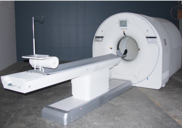
Model mock up of whole body PET scanner
HCB News: Do you ever hear from skeptics who question the clinical value of your research? What do you tell them?
B & C: Absolutely! I would say that when we started, probably three-quarters of our colleagues in the field questioned us on the value of this. However, as we have kept making the arguments, that number has probably gone down to about 20 percent, and there are now multiple sites across the world that have expressed an interest in obtaining a total-body PET scanner.
Actually, we welcome skepticism. It ensures that we stay on track with our arguments and helps prevent us from engaging in sloppy thinking. The biggest argument we hear is that the device will be just too expensive. But when all the installation requirements are factored in, this device won't be very much more expensive than a very-high-field (e.g. 7 tesla) MRI scanner, and we believe it opens up many more avenues of research. We are looking forward to using this scanner to try to answer a whole range of questions that we could not hope to look at before, and to finding out who is right! Also, as with all new technologies, initial costs often are high, but if we can demonstrate clear benefit that, in turn, generates demand, then there will be pressures and opportunities to reduce cost.